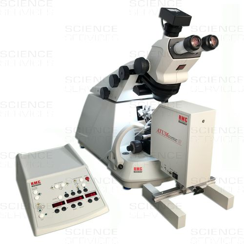
ATUMtome – Automated Tape Collecting Ultramicrotome
R-ATUMtome
The ATUMtome by RMC, a tape collecting ultramicrotome with automated section collector. Initially developed at Harvard University in the lab of Jeff Lichtman. The ATUMtome is a unique advanced system for ultrathin sectioning of resin-embedded specimens and collecting sections on tape for SEM imaging and subsequent 3-D reconstruction (Array Tomography).
Product Details
Description
Features and Benefits of the ATUMtome
- Collects hundreds to thousands of sections on a continuous tape
- Convenient section collection even for small series
- Non-destructive system compared to en-bloc techniques (FIB-SEM, SB-SEM)
- Data collection is decoupled from sectioning allowing multiple imaging runs with various resolutions
- Sample storage for years on wafers for further processing, post-staining, immunogold labeling, correlative microscopy
- Sample viability is determined earlier in the process compared to en-bloc techniques
- Uses standard sample preparation techniques and resins compared to en-bloc techniques
- Section thickness range from 30nm to 5µm (even less possible)
- Low costs compared to alternative 3-D EM data collection approaches (FIB-SEM, SBSEM)
Working Principle of the ATUMtome
The ATUMtome is a unique ultramicrotome for collecting sections on a continuous tape. Serial sections of resin-embedded specimens are cut with a diamond knife. As the sections float off on the water surface in the knife trough, they are picked up by the moving tape of the ATUM. The long rolls of tape are cut into shorter lengths and mounted on silicon wafers.
The populated wafers are now ready for imaging with a scanning electron microscope (SEM). Sections can be stored and archived in wafer-libraries and processed again at any time in the future.
Papers & Publications
This is a list of papers and other publications that mention the use of the ATUMtome. The latest papers, videos and puplications you can find here.
July 2020 — Multiscale Imaging with Photons, Electrons and Ions (Neuromethods 155).
https://www.ebookee.com/Volume-Microscopy-Multiscale-Imaging-with-Photons-Electrons-and-Ions-Neuromethods
June 2020 — Multiscale ATUM-FIB Microscopy Enables Targeted Ultrastructural Analysis at Isotropic Resolution (Cell).
https://www.cell.com/iscience/fulltext/
March 2020 — Serial section Raman tomography with 10 times higher depth resolution than confocal Raman microscopy (Raman Spectroscopy).
https://www.onlinelibrary.wiley.com/doi/full/
October 2019 — Presynaptic Mitochondria Volume and Abundance Increase during Development of a High-Fidelity Synapse (JNeurosci).
https://www.jneurosci.org/content/39/41/7994?iss=41
October 2019 — Serial-section electron microscopy using automated tape-collecting ultramicrotome (ATUM) (Elsevier).
https://www.sciencedirect.com/science/article/pii/
February 2019 — Lateral Inhibition in Cell Specification Mediated by Mechanical Signals Modulating TAZ Activity (Cell).
https://www.cell.com/cell/fulltext
January 2018 — A carbon nanotube tape for serial-section electron microscopy of brain ultrastructure (Nature Communications). (Yoshiyuki Kubota, Jaerin Sohn, Sayuri Hatada, Meike Schurr, Jakob Straehle, Anjali Gour, Ralph Neujahr, Takafumi Miki, Shawn Mikula & Yasuo Kawaguchi) The paper discribes a new tape material and optimized workflows to enable high-resolution volume electron microscopy for brain ultrastructure analysis. https://www.nature.com/articles/s41467-017-02768-7
July 7, 2016 — Reconstruction of genetically identified neurons imaged by serial-section electron microscopy (eLife). (Maximilian Joesch, David Mankus, Masahito Yamagata, Ali Shahbazi, Richard Schalek, Adi Suissa-Peleg, Markus Meister, Jeff W Lichtman, Walter J Scheirer, and Joshua R Sanes) Discusses targeted reconstruction of specific neuronal types as an alternative strategy for complete circuit reconstruction. http://www.ncbi.nlm.nih.gov/pmc/articles/PMC4959841/.
June 2016 — High-Throughput, High-Resolution Mapping of Protein Localization in Mammalian Brain by In Vivo Genome Editing (Cell). — (Takayasu Mikuni, Jun Nishiyama, Ye Sun, Naomi Kamasawa, and Ryohei Yasuda) Paper discusses a scalable and high-throughput method to identify precise subcellular localization of endogenous proteins essential for integrative understanding of a cell at the molecular level. (Register for full access) http://www.cell.com/cell/abstract/S0092-8674(16)30489-5.
August 2015 — On the Road to Large Volumes in LM and SEM: New Tools for Array Tomography (Microscopy & Microanalysis). (Irene Wacker, Waldmar Spomer, Andreas Hofmann, Ulrich Gengenbach, Marlene Thaler, Len Ness, Pat Brey and Rasmus R. Schroder) Paper discusses the advantage of array tomography over serial blockface methods of reconstructing large volumes of samples at ultra-structural resolution. (Pay for full access) http://journals.cambridge.org/action/displayAbstract?fromPage=online&aid=9918154
August 2015 — Advanced Substrate Holder and Multi-Axis Tool for Ultramicrotomy (Microscopy & Microanalysis). (Waldemar Spomer, Andreas Hofmann, Irene Wacker, Lenn Ness, Pat Brey, Rasmus . Schroder and Ulrich Gengenbach). A novel, multi-axis device is now being developed to support ultramicotome users in transferring sections onto a solid substrate. (Pay for full access.) http://journals.cambridge.org/action/displayAbstract?fromPage=online&aid=9919140#
July 30, 2015 — Cell, Saturated Reconstruction of a Volume of Neocortex (Lichtman Lab). This paper describes a suite of automated technologies designed to probe the structure of neural tissue at nanometer resolution, including the ATUMtome. This approach is used to generate a “saturated” reconstruction of a 1500 cubic micron sub-volume of mouse neocortex. Several related links provided below:
Lab’s Intro: https://www.mcb.harvard.edu/mcb/news/news-detail/3821/reconstructing-the-brains-wiring-diagram-lichtman-lab/
Related video: http://lichtmanlab.fas.harvard.edu/files/lichtman/files/lichtman_cell_2015.mp4
April 13, 2015 — Nature Methods, High-resolution whole-brain staining for electron microscopic circuit reconstruction ( Shawn Mikula & Winfried Denk). Currently only electron microscopy provides the resolution necessary to reconstruct neuronal circuits completely and with single-synapse resolution … “Our results suggest that the BROPA method can produce a preparation suitable for the reconstruction of neural circuits spanning an entire mouse brain.” Full article may require subscription. http://www.nature.com/nmeth/journal/v12/n6/full/nmeth.3361.html
February 2015 — Microscopy, Oxford Journals, New Developments in Electron Microscopy for Serial Image Acquisition of Neuronal Profiles (Yoshiyuki Kubota). Covers recent developments in electron microscopy that largely automate the continuous acquisition of serial electron micrographs (EMGs). http://jmicro.oxfordjournals.org/content/64/1/27.full?sid=c6cd6532-de6e-49af-9fcb-878aa8e59cd7
October 23, 2014 — Oxford Journals, Review: Mission (im)possible — mapping the brain becomes an reality (Anna Lena Eberle, Olaf Selchow, Marlene Thaler, Dirk Zeidler and Robert Kirmse) http://jmicro.oxfordjournals.org/content/64/1/45.full.pdf?keytype=ref&ijkey=vzgj1nAZfGBItHz
October 20, 2014 — Microscopy & Microanalysis, Positional correlative anatomy of invertebrate model organisms increases efficiency of TEM data production. (Kolotuev) Includes some interesting approaches to specimen preparation. http://www.ncbi.nlm.nih.gov/pubmed/25180638
October 3, 2014 – Biorxiv.org, Conneconomics: The Economics of Dense, Large-Scale, High-Resolution Neural Connectomics (Adam H. Marblestone,
Evan R. Daugharthy, Reza Kalhor, Ian D. Peikon, Justus M. Kebschull, Seth L. Shipman, Yuriy Mishchenko, Jehyuk Lee, David A. Dalrymple, Bradley M. Zamft, Konrad P. Kording, Edward S. Boyden, Anthony M. Zador and George M. Church) http://biorxiv.org/content/biorxiv/early/2014/04/20/001214.full.pdf
June 27, 2014 – Frontiers in Neural Circuits, Imaging ATUM ultrathin section libraries with WaferMapper: a multi-scale approach to EM reconstruction of neural circuits ( Kenneth J. Hayworth, Josh L. Morgan, Richard Schalek, Daniel R. Berger,David G. C. Hildebrand and Jeff W. Lichtman) http://www.ncbi.nlm.nih.gov/pmc/articles/PMC4073626/
July 18, 2013 — Cell, Stacked endoplasmic reticulum sheets are connected by helicoidal membrane motifs (Mark Terasaki, Tom Shemesh, Narayanan Kasthuri,Robin W. Klemm,Richard Schalek, Kenneth J. Hayworth,Arthur R. Hand,Maya Yankova,Greg Huber,Jeff W. Lichtman,Tom A. Rapoport,and Michael M. Kozlov) http://www.cell.com/cell/abstract/S0092-8674%2813%2900770-8?_returnURL=http%3A%2%2Flinkinghub.elsevier.com%2Fretrieve%2Fpii%2FS0092867413007708%3Fshowall%3Dtrue
2014 – University of Heidelberg, Dissertation: Learning-based Segmentation for Connectomics (Diplom-Physiker Thorben Kroger) http://archiv.ub.uni-heidelberg.de/volltextserver/16834/1/diss.pdf
2014 – University of California at Berkley, Optimization-Based Artifact Correction for Electron Microscopy Image Stacks (Samaneh Azadi,
Jeremy Maitin-Shepard and Pieter Abbeel) http://link.springer.com/chapter/10.1007/978-3-319-10605-2_15#page-1
May 2013 – Nature Methods, Focus on Mapping the Brain (Erika Pastrana) http://www.nature.com/nmeth/journal/v10/n6/full/nmeth.2509.html
2013 – Journal of Cell Science, Commentary: Is EM dead? (Graham Knott and Christel Genoud) http://jcs.biologists.org/content/126/20/4545.full
July 2012 — Microscopy & Analysis, ATUM-based SEM for High-Speed Large-Volume Biological Reconstructions ($$) (R. Schalek, A. Wilson, J. Lichtman, M. Josh, N. Kasthuri, D. Berger, S. Seung, P. Anger, K. Hayworth and D. Aderhold) http://journals.cambridge.org/action/displayAbstract?fromPage=online&aid=8758526
April 16, 2012 – PLOS One, Serial Section Scanning Electron Microscopy (S3EM) on Silicon Wafers for Ultra-Structural Volume Imaging of Cells and Tissues (Heinz Horstmann, Christoph Körber, Kurt Sätzler, Daniel Aydin, Thomas Kune) http://www.plosone.org/article/info%3Adoi%2F10.1371%2Fjournal.pone.0035172
January 12, 2012 — Nature Protocols, FESEM images of adult Drosophila brain ultrathin section mounted on Kapton tape. ( Juan Carlos Tapia, Narayanan Kasthuri, Kenneth J Hayworth, Richard Schalek, Jeff W Lichtman, Stephen J Smith & JoAnn Buchanan) http://www.nature.com/nprot/journal/v7/n2/fig_tab/nprot.2011.439_F7.html
November 24, 2011 — Science Direct,Volume electron microscopy for neuronal circuit reconstruction (Kevin L Briggman and Davi D Bock) https://archive.org/stream/pdfy-jFNzCxKoDhqFxdNq/2012-briggman_djvu.txt
2011 — Microscopy & Microanalysis, Development of High Throughput, High Resolution 3D Reconstruction of Large Volume Biological Tissue Using Automated Tape Collection Ultramicrotomy and Scanning Electron Microscopy (R Schalek, N Kasthuri, K Hayworth, D Berger, J Tapia, J Morgan, S Turaga, E Fagerholm, H Seung and J Lichtman) http://journals.cambridge.org/action/displayAbstract?fromPage=online&aid=8396995&fileId=S1431927611005708
ZEISS offers a dedicated soft- and hardware package that allows researchers to image the large number of ATUMtome serial sections in an automated way. ZEISS Atlas 5 Array Tomography is available for all ZEISS scanning electron microscopes.
More Information
| Manufacturer |
RMC Boeckeler
|
|---|---|
| Dimensions | 124,5 x 91,4 x 137,2cm |
| Electrical Power | 110 - 240 VAC 50/60 Hz |
| Packing Unit | each |
| Weight | 361 kg) |
Downloads

You may be also interested in following products
-
Starting at
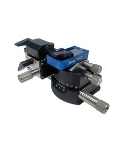
The ASH2 is used to efficiently collect and align hundreds of serial sections onto a variety of substrates for subsequent imaging in LM and SEM. The advanced substrate holder ASH2 quickly mounts onto the knife stage of RMC PowerTome ultramicrotomes.
Starting at

Who Viewed This Also Viewed