A dye for FIRM – Fluorophore-Infiltrated Resin Microscopy
Product Details
Description
Functional Principle:
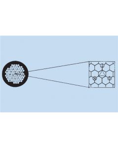
The key principle is the infiltration of thin resin sections with a special fluorophore. Upon fluorescence illumination, the resin appears brightly fluorescent revealing cells and tissue structures in a negative relief (dark).
- Works with Epoxy and Acrylic type resins
- A unique way of staining - fluorescent dye infiltrates the resin without staining the tissue revealing structures in a negative relief
- Extremely high contrast and resolution
- Fast preliminary analysis of samples prior to EM analysis
- Also ideal for material and food science analytical microscopy
Application Protocol:
- Simply mount your sections on glass slides
- Air dry, and overlay with 100µl of FIRM for 30 seconds
- Rinse and coverslip with water or aqueous mounting medium
- View in Rhodamine channel of a standard wide-field Fluorescence Microscope
Human anterior pituitary, FIRM, rhodamine channel. Note that the fluorophore illuminates the LR White resin, providing an image of tissue structure primarily in negative relief. Bar, 100µm. All subsequent FIRM images are shown in monochrome.

Human anterior pituitary, formalin-fixed autopsy specimen, comparison of H&E paraffin section (left) with FIRM (right). 20x dry objective used for both. Scale bar, 20µm.

Human anterior pituitary, FIRM image taken with 150X glycerin objective. Individual secretory granules are easily visualized, ranging from 150- 350nm in diameter. Scale bar, 5µm.

Correlative FIRM/fluorescent lectin labeling of human pituitary. A single LR White thin section was labeled first with Concanavalin AAlexa-488 (B) imaged, then stained for FIRM imaging (A). Densely granulated cells (arrows in A) are strongly labeled with Con-A (arrows in B). Dark structures resembling large lysosomes or residual bodies (arrowheads in A) are also strongly labeled by Con-A, consistent with reports in the literature.

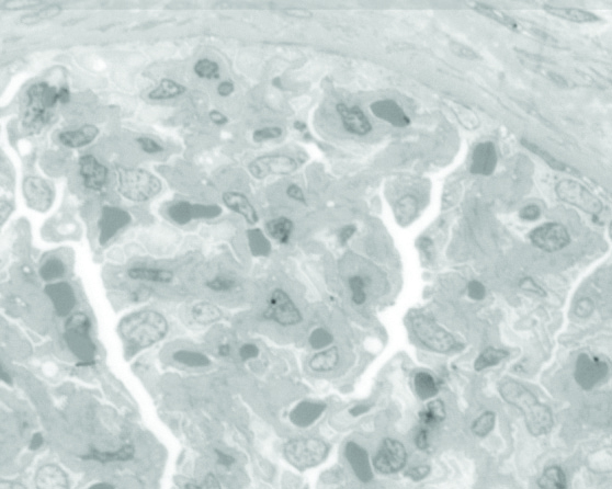
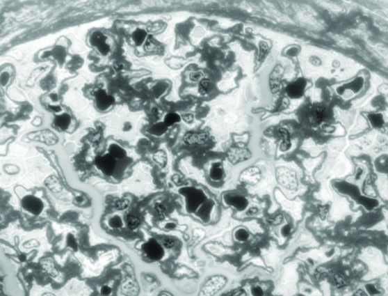
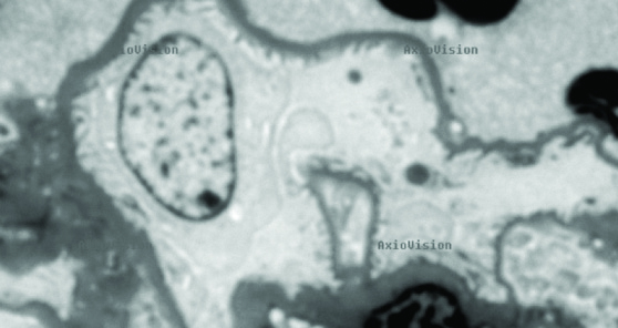
Comparison of toluidine blue staining (top) to FIRM (middle) on near adjacent sections of human kidney. Note enhanced contrast, especially of glomerular basement membranes in FIRM image. High power FIRM image (bottom) of glomerulus. Note resolution of podocyte foot processes (arrows). Scale bars in A, B, 20µm, in C, 5µm.


Who Viewed This Also Viewed