Interview with Claudia Mayrhofer - Graz Centre for Electron Microscopy (ZFE)
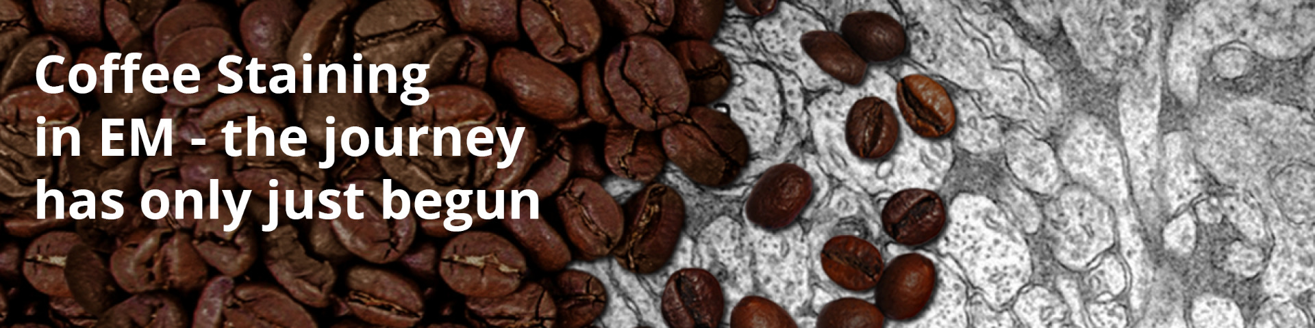
With their publication `Coffee – a ubiquitous substitute for uranyl acetate in staining of biological ultrathin sections for electron microscopy studies’ researchers led by Claudia Mayrhofer have caused quite a stir in various newspapers. We conducted the following interview with Claudia to find out more.
Claudia Mayrhofer: After completing a technical high school education (HTL) in electrical engineering and beginning a degree in chemistry, Claudia Mayrhofer worked in the paper industry before joining the Technical University of Graz in 2003, where she began working in the field of electron microscopy. There, she discovered her passion for ultramicrotomy, which she continues to pursue to this day.
How did you come up with the idea of using coffee as a staining agent?
The initiation was the ban on uranyl acetate in our lab in 2019. We could no longer order it; storage became increasingly complicated, and disposal costs increased tremendously. I still had some stock left, but not enough to last forever.
At first, we used platinum blue, but it’s almost more toxic than uranyl acetate, and I didn’t want to expose my students and colleagues to that.
Then we thought about OTE (Oolong Tea Extract), but during the COVID-19 pandemic, we couldn’t get hold of the high-purity extract.
One day, I was sitting in the lab, looking at my colleagues’ dried coffee cups and thought: If tea works, why not coffee? Both contain similar tannins. So, I decided to use coffee grounds to stain algae from my aquarium — and it worked right away!
The first test was so successful that I repeated it with freshly brewed coffee — and the results were even better. That’s how it all started!
How long did you work on the coffee-contrast project?
It was quite a long process! After the initial successful tests with algae, we tried to validate the method scientifically. Our Master’s student, Robert Zandonella, developed a MATLAB program to quantitatively assess contrast quality on mitochondria since they are particularly suitable for measurement. The focus was on the application and evaluation of various alternative staining agents for comparison. The classic contrast staining with uranyl acetate and lead citrate according to Reynolds served as the reference method.
For each mitochondrion, ten points were analyzed to eliminate subjective bias. That step was crucial because a visually good image doesn’t necessarily mean reproducibility. The Master’s thesis took quite some time, during which we systematically compared the relevant compounds.
The publication process turned out to be unexpectedly tough, partly due to the unconventional nature of the method and the initial difficulty finding a second reviewer. But now the paper is finally out, and the response is far greater than I had ever expected.
Which compound in coffee is responsible for the contrast?
After the initial successful staining, our first thought was to test the coffee for heavy metals — unlikely, but you never know how coffee is treated. Once this test came back negative, we analyzed the coffee components and identified chlorogenic acid as the key factor responsible for the good contrast.
Which coffee bean or roast works best?
That’s a science in itself! We tested different beans and roast levels — and yes, there are differences. Unroasted green coffee worked best but is extremely difficult to handle: the beans must be ground, boiled, and filtered, and the extract is so inhomogeneous that every syringe filter clogs immediately. Not a practical option for daily work.
Standard Arabica coffee varies too much in roast, strength, and brewing conditions. For scientific purposes, you need consistent parameters. Therefore, we use coffee extract only for preliminary trials.
How reproducible is coffee-based staining?
Here lies the big difference between coffee extract and pure chlorogenic acid. The extract is a great quick solution, but the results are highly variable. Depending on bean type, roast, or even brewing temperature, the extract’s composition changes. That’s a serious issue for reproducibility in scientific research.
For this reason, we focused on chlorogenic acid — it’s commercially available, stable, and produces reproducible results. Even better: samples contrasted with chlorogenic acid remained perfectly intact after 1.5 years, while those stained with uranyl acetate or coffee extract showed precipitates.
What are the advantages of coffee/chlorogenic acid compared to tea?
Tea or OTE also works, but coffee extract and chlorogenic acid produce sharper contrasts. OTE, which we later obtained, showed pronounced precipitates that we couldn’t eliminate — unlike coffee.
Do you regularly use coffee extract and chlorogenic acid in your facility?
Absolutely! For quick tests or preliminary staining, we use coffee extract — it’s inexpensive and works well. For scientific projects or long-term studies, we rely on chlorogenic acid because of its reproducibility, especially for established samples such as algae or yeast.
With new specimen types, we always test both — but chlorogenic acid almost always wins. Best of all, the students love it, since they can work without hazardous chemicals!
Which specimens work, and which don’t?
So far, we’ve successfully used chlorogenic acid on algae, yeast, rat kidney, mouse liver, and zebrafish. The contrast is particularly striking in biological samples such as algae or yeast. Negative staining, on the other hand, has worked less well.
For polymers or other plastics, I see less potential, as uranyl acetate is rarely used there anyway. Unfortunately, we’ve only been able to work to a limited extent with native animal or human tissues because our institute lacks the necessary authorization. That’s a pity, since chlorogenic acid shows great potential in biological applications. We hope to collaborate with other labs that can prepare such samples — perhaps through our EM lab network!
What are the differences compared to uranyl acetate?
The most significant difference is sample cleanliness. Uranyl acetate often leaves precipitates after staining, even with thorough washing — chlorogenic acid doesn’t. Moreover, chlorogenic-acid-stained samples exhibit better long-term stability: after months of storage, they remained flawless, whereas uranyl-acetate-stained samples already showed contamination.
In addition, I find chlorogenic acid produces stronger contrast — mitochondrial contours and other structures appear sharper.
And the biggest advantage: chlorogenic acid is non-toxic and genuinely makes life easier — and apparently, without compromising quality.
Is this the end of development, or what’s next?
Definitely not the end! On the contrary — I want to further develop the method for negative staining. We’ve tried that a few times, but with mixed success so far. There’s still a lot of room for optimization.
For that, we need more tissue samples and, above all, partner laboratories that can provide samples or conduct parallel experiments. My dream is a large network of laboratories that jointly test, compare, and establish the method. Maybe something will emerge through the community and upcoming meetings.
The journey has only just begun — and I’m excited to see where it leads!
You also run ultramicrotomy workshops — how can people learn more? And are there courses on coffee staining?
At ZFE (Zentrum für Elektonenmikroskopie), we offer customized ultramicrotomy courses for both internal and external participants. In the past, these were often group courses with ten or more participants, but because of the diversity of samples in biological and materials research, we realized that such group formats weren’t very efficient.
Now, I provide tailored training: either participants come to us, or I go to their facilities and work directly on their instruments — much more effective!
There aren’t any specific coffee-staining courses yet, but I’m open to inquiries. A ring trial with other labs would be particularly exciting to further test the method. Anyone interested is welcome to contact me — we’ll definitely find a solution!
By the way, everyone who books a course with me receives a copy of the new ultramicrotomy book “Ultramicrotomy in Materials and Life Sciences” — which includes a chapter co-authored by Helmut Gnägi and myself!