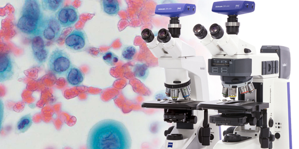
Microscopy Solutions for Cytology and Cytopathology
Microscopy applications for your laboratory
Created by Carl Zeiss Microscopy GmbH
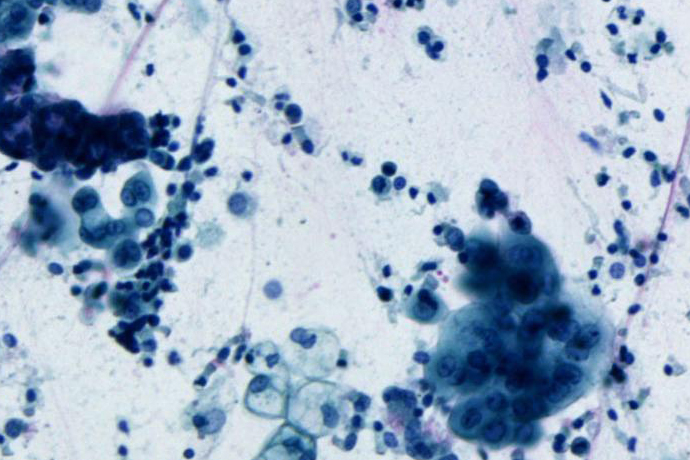
Prominent examples for exfoliative cytology are the aforementioned Pap smear, where cells are scraped from the cervix with a cervical spatula, or cells that are harvested from bodily fluids such as blood and urine, pleural and pericardial effusion, or ascites from the peritoneum. In intervention or fine-needle aspiration cytology (FNAC) the pathologist uses a small needle to collect cells in various body organs for diagnostic analysis in gynecologic, lymph node, thyroid, breast, liver, lung, kidney or pancreas cytology. These cytological screenings are most commonly used to search for cells going through abnormal changes called hyperplasia and dysplasia that may become cancer cells.
Microscopy Solutions for Cytology and Cytopathology
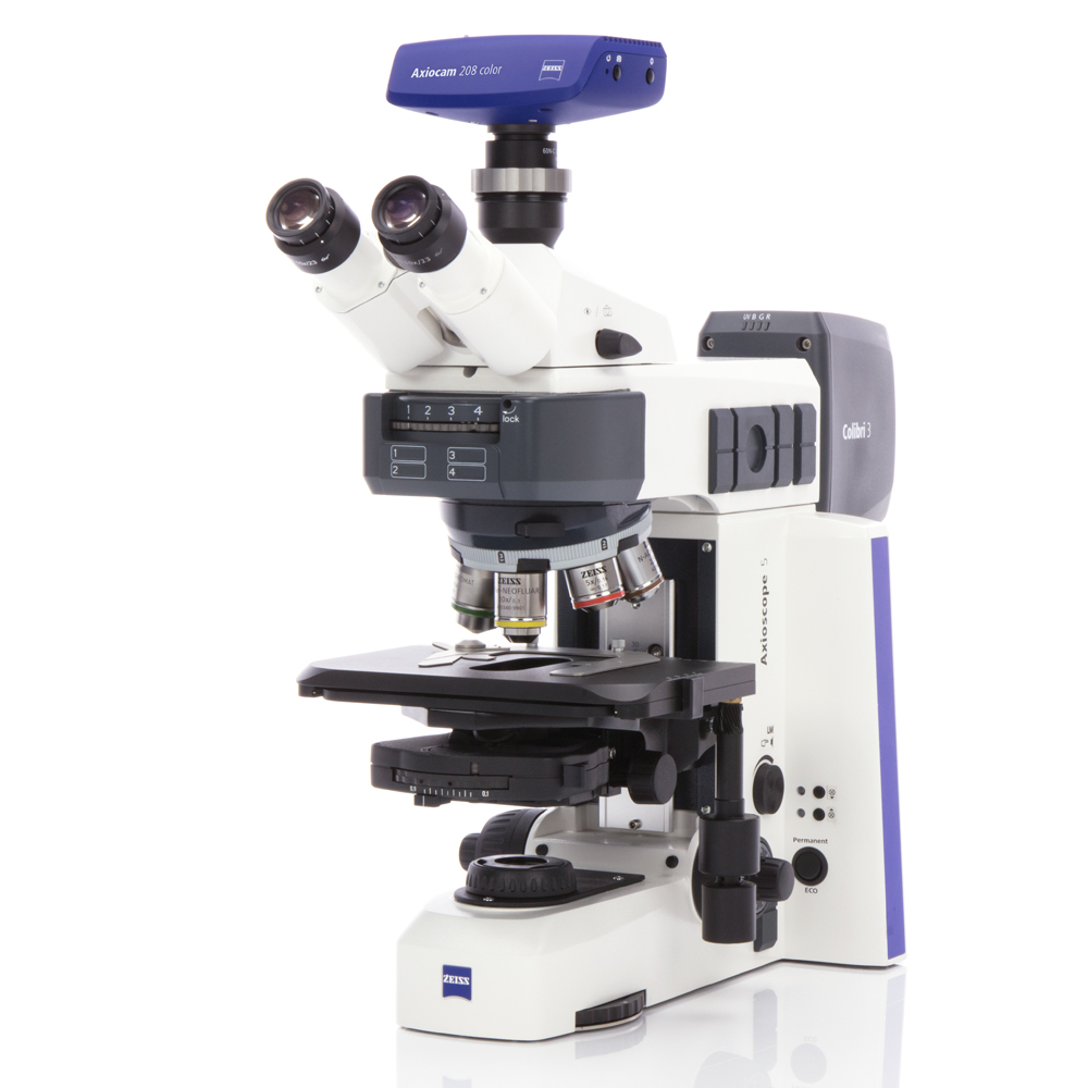
Microscope Requirements
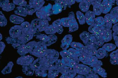
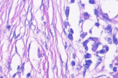
A very good cellular differentiation and clearly visible nuclear details are absolute prerequisites in cytology for carcinoma and tumor cell diagnosis. Cytologists and pathologists rely on crystal-clear images of their samples with the highest color fidelity in brightfield, phase contrast, DIC, or fluorescence. While cytological stains such as Papanicolaou’s (PAP stain), Giemsa, or Romanowski-type result in specific staining of cellular features, it is the optical quality of the microscope, the fidelity of the attached camera for digital documentation, and the ergonomic design of the instrument that can make all the difference when screening patient samples.