Types of Microscopy
Standard bright-field microscopy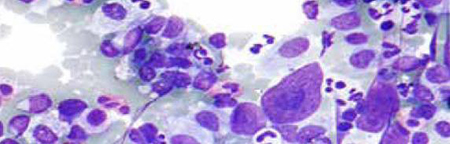
Standard bright-field microscopes are used for daily laboratory routine in research and diagnostics for simple, standard applications that require no special equipment. Therefore simple optical systems and lenses are applied.
Phase contrast microscopy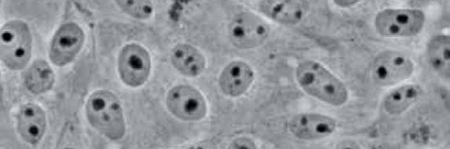
Phase contrast microscopy is a contrast-enhancing technique to visualize structures difficult to detect with bright-field microscopy due to a lack of contrast, without the need of a staining. When penetrating a medium, light propagates with different speeds depending on the refractive index of the medium. This leads to phase differences, which are converted to differences in brightness by the microscope using phase rings. Areas of application of phase contrast microscopes are mainly the observation of living biological samples in order to resolve fine structures with high contrast.
Fluorescence microscopy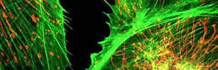
The physical principle of fluorescence is used to selectively visualize and localize defined fluorescent structures, while non-fluorescent structures remain dark in order to obtain a high image contrast. For this purpose, fluorescent dyes (fluorochromes) are used with specific excitation and emission filters installed in the optical path of the microscope. A wide range of fluorescent dyes with different colors are available, which are used in molecular biological, biomedical and clinical research. For example, in immunohistochemistry, fluorescence-in-situ-hybridization and for visualization of cells or cellular components in living/fixed specimens.
Dark field microscopy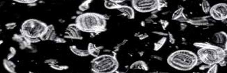
A dark field microscope produces a contrast-enhanced image by indirect illumination of the specimen, thereby also unstained specimens can be displayed with high contrast. Using this technique, direct light is bypassing the objective, only light scattered by the specimen enters the objective. As a result, the background appears dark or black, only the specimen is illuminated and even small structures can be visualized with high contrast. Dark field microscopy in biology and medicine is especially used for transparent and low contrast specimens, for example, studying blood, small animals or micro-particles in material science.
Polarization microscopy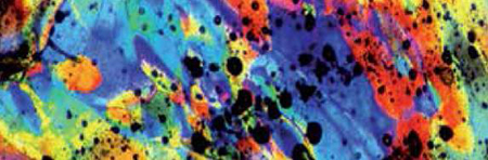
Polarization microscopy is used for the analysis of optically anisotropic samples. The primary objective of polarization microscopy is not magnification of an object, but rather the analysis of optical properties such as refractive index or birefringence for sample analysis. The method is mainly used in mineralogy and in industry for testing plastics or mineral building materials in order to gain insights into their composition.
Metallurgical microscopy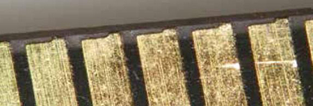
A metallurgical microscope is a special version of a standard light microscope for the study of materials, such as Metals, plastics, ceramics and others. Since materials are usually not transparent solid structures, metallurgical microscopes often have an upright light unit. Moreover, this type of microscope is characterized by extensive magnification, e.g. for detailed investigations of surface structures. Application areas of metallurgical microscopy are industry, material science and research.
Inverted microscopy
An inverted microscope is an upside-down standard light microscope. This type of microscope is characterized by locating the objective underneath the stage and pointing upwards to the specimen. Inverted microscopes are mainly used for live cell analysis of cell cultures growing in culture medium. Culture dishes are not only available with standard coverslip bottoms (thickness 0.17mm) but also in various material and bottom thicknesses. Therefore, for some models special objectives are available that can correct for different bottom glass thicknesses. In addition, inverse microscopes are also used for studies of thicker specimens. Inverted microscopes are also available as dark field, polarization or fluorescence microscopes.