+49 (089) 18 93 668 0 - Info@ScienceServices.de   We are here for you! Call us or request a call back here.
Language

UranyLess: Applications

Plant Tissue

Preparation of the sample using the following protocol:

  • Glutaraldehyde Fixation Classic - Osmium - Included in Epon
  • Contrast the UranyLess monitoring Lead Citrate
UranyLess - Plant Tissue UranyLess - Plant
Plant Leaf. Photo: Jeannine
Lherminier (INRA - Dijon)
Plant Leaf. Photo: Jeannine
Lherminier (INRA - Dijon)


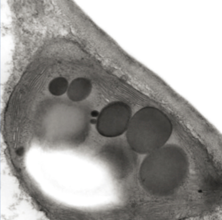
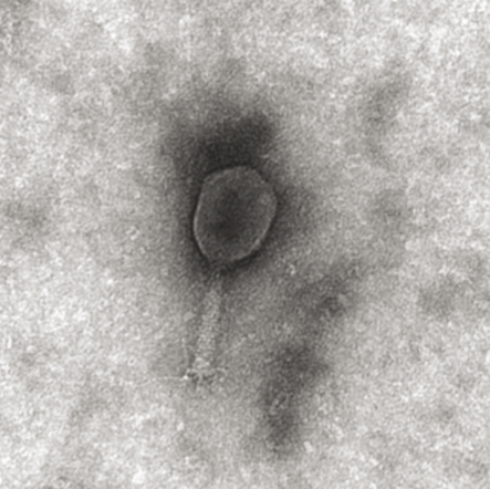
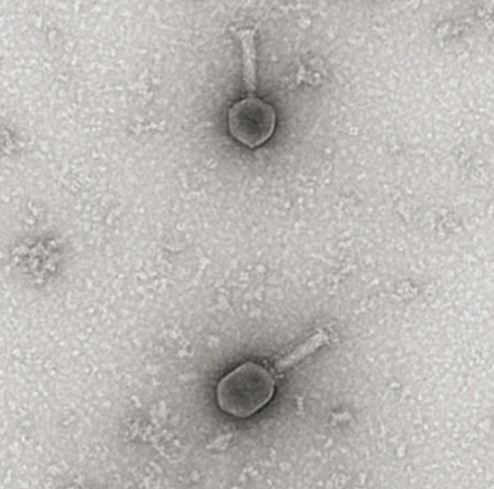
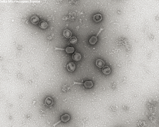
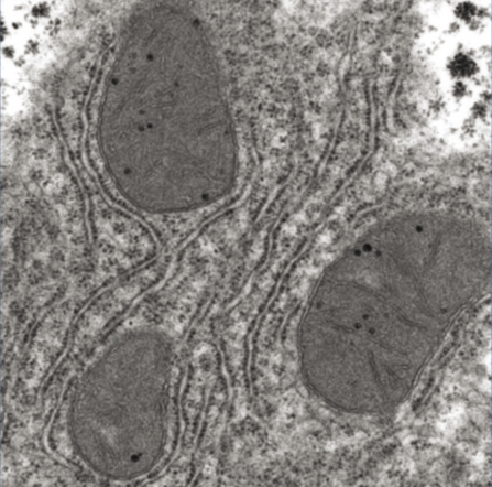
Phage T6

Preparation of the sample using the following protocol:
  • Staggering Phage T6 on a G300-Cu grid Covered with a Carbon Formvar Film. Ionization 1 minute
UranyLess - Phage T6 UranyLess - Phage T6 UranyLess - Phage T6 UranyLess - Phage T6
Phage. Photo: Nacer
Benmeradi (R & D - DeltaMicroscopies-France)
Phage. Photo: Nacer
Benmeradi (R & D - DeltaMicroscopies-France)
Phage. Photo: Nacer
Benmeradi (R & D - DeltaMicroscopies-France)
Phage. Photo: Nacer
Benmeradi (R & D - DeltaMicroscopies-France)


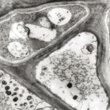
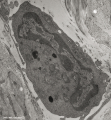
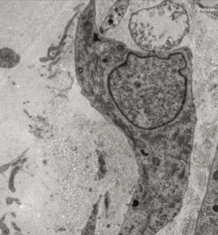
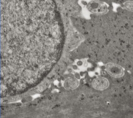
Liver Mouse and Gerbil Sahara

Preparation of the sample using the following protocol:
  • Classic-Glutaraldehyde Fixation PFA, Osmium, Epon
  • Ultrafine Cup - Contrast UranyLess Lead - Citrate
UranyLess - Liver Mouse and Gerbil Sahara
Hepatocyte - Perinuclear Region.
Photo: Nacer Benmeradi
(R & D - DeltaMicroscopies-France)


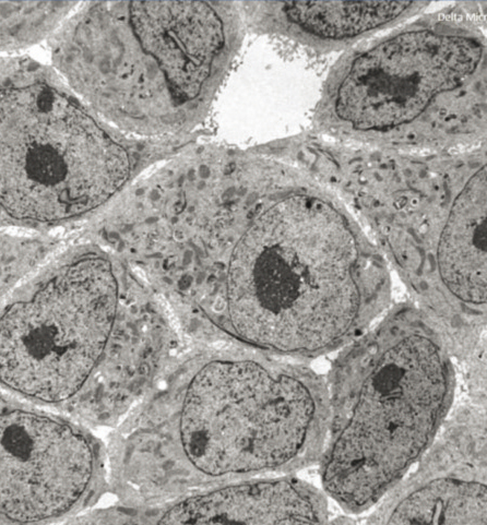
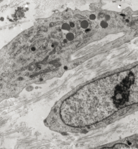
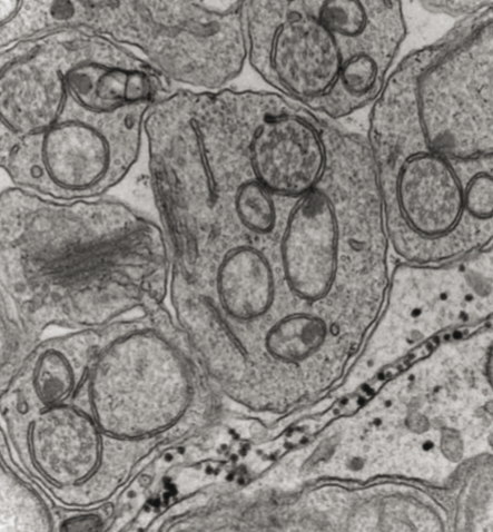
Culture Cells, Preparation of the sample using the following protocol:

  • Classic-Glutaraldehyde Fixation PFA, Osmium, Epon
  • Ultrafine Cup - Contrast UranyLess lead -citrate
UranyLess - Culture Cells UranyLess - Culture Cells UranyLess - Culture Cells UranyLess - Culture Cells UranyLess - Culture Cells
Culture Cell. Photo: Nacer
Benmeradi(R & D - DeltaMicroscopies-France)
Culture Cell. Photo: Nacer
Benmeradi(R & D - DeltaMicroscopies-France)
Culture Cell. Photo: Nacer
Benmeradi(R & D - DeltaMicroscopies-France)
Culture Cell. Photo: Nacer
Benmeradi(R & D - DeltaMicroscopies-France)
Culture Cell. Photo: Nacer
Benmeradi(R & D - DeltaMicroscopies-France)


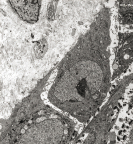
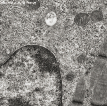
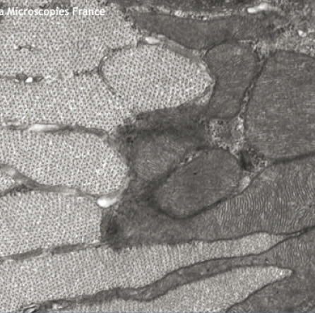
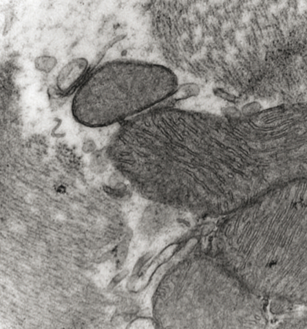
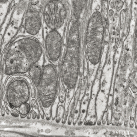
Muscle - Nerve - Mice

Preparation of the sample using the following protocol:

  • Classic-Glutaraldehyde Fixation PFA, Osmium, Epon
  • Ultrafine Cup - UranyLess Contrast 1 minute followed lead Citrate 1 minute
UranyLess - Muscle, Nerve, Mice UranyLess - Muscle, Nerve, Mice UranyLess - Muscle, Nerve, Mice UranyLess - Muscle, Nerve, Mice
 Longitudinal Section of Mouse
Skeletal Muscle - Nerve Cup
(dense area myelin sheath).
Photo: Nacer Benmeradi
(R & D - DeltaMicroscopies-France)
Detailed View of Myocytes.
Photo: Nacer Benmeradi
(R & D - DeltaMicroscopies-
France)
Cross Section of Muscle
Fibers - Mitochondria.
Photo: Nacer Benmeradi
(R & D - DeltaMicroscopies-
France)
Myocyte. Photo: Nacer
Benmeradi (R & D -
DeltaMicroscopies-France)


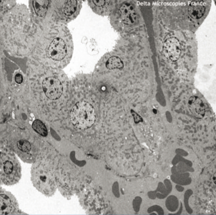
PLC Contrast Leica EM Stain

Preparation of the sample using the following protocol:

  • Classic-Glutaraldehyde Fixation PFA, Osmium, Epon
  • Ultrafine Cup - contrast Uranyless Lead -Citrate

Chantal Cazevieille CRIC / IURC INSERM Montpellier tested aqueous UranyLess in the Leica brand grid contrast controller on different tissues, Drosophila heart atrium, retina, cochlea and ileum (Gut). The tissues were fixed according to the standard protocol 2.5% Glutaraldehyde in PHEM buffer, the post fixation in 0.5% osmium in 0.8% potassium ferrocyanide in RT for 2 hours. The sections are collected on single-hole or 200 mesh grids.
The treatment of the grids is UranyLess 7mn lead citrate followed 7 minutes. We present here some images made by Hitachi transmission electron microscope with a digital camera AMT. You will notice that the combined action of potassium ferrocyanide and UranyLess reveal a marked way the cyto-membranes in the ileum.

UranyLess - PLC Contrast UranyLess - PLC Contrast UranyLess - PLC Contrast UranyLess - PLC Contrast
Spermatides Drosophile.
Photo: Chantal Cazevieille
Montpellier
Heart Headset.
Photo: Chantal Cazevieille
CRIC / IURC la'INSERM Montpellier
(R & D - DeltaMicroscopies-France)
Drosophila. Photo: Chantal
Cazevieille CRIC / IURC
la'INSERM Montpellier
Drosophila. Photo: Chantal
Cazevieille CRIC / IURC
la'INSERM Montpellier


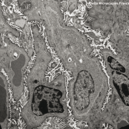
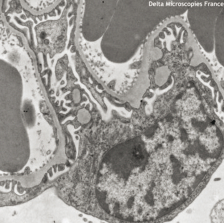
Mouse Kidney

Preparation of the sample using the following protocol:

  • Classic-Glutaraldehyde Fixation PFA, Osmium, Epon
  • Ultrafine Cup - UranyLess Contrast 1 minute followed lead Citrate 1 minute
UranyLess - Mouse Kidney UranyLess - Mouse Kidney UranyLess - Mouse Kidney UranyLess - Mouse Kidney
Mouse Kidney.
Photo: Nacer Benmeradi
(R & D - DeltaMicroscopies-
France)
Basal Invaginations -
Hémidesmosome -
Basal Lamina: Increase the
Exchange Surface - Kidney.
Photo: Nacer Benmeradi
(R & D - DeltaMicroscopies-
France)
Glomerular area - Podocytes -
Stalks - Kidney. Photo:
Nacer Benmeradi
(R & D - DeltaMicroscopies-France)
Podocyte - Pedicels - Kidney.
Photo: Nacer Benmeradi
(R & D - DeltaMicroscopies-France)


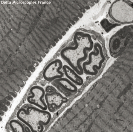
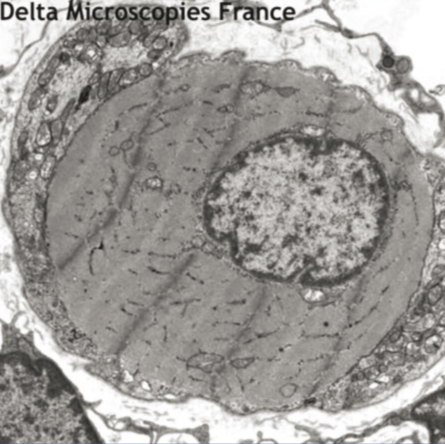
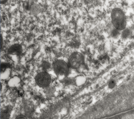
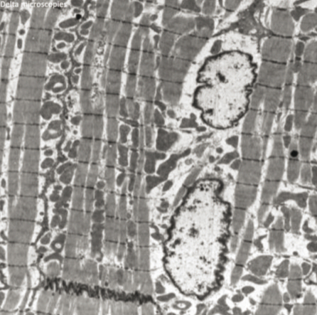
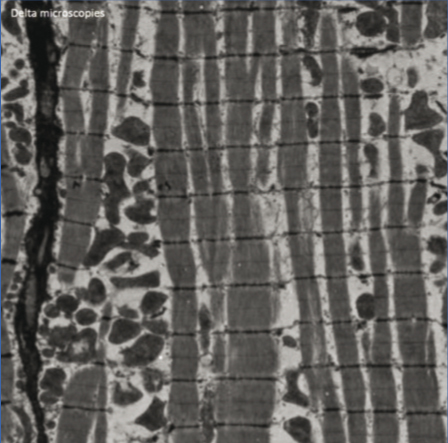
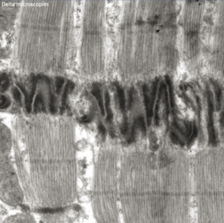
Mouse Cardiac Muscle

Preparation of the sample using the following protocol:

  • Classic - Fixing Glutaraldehyde, Osmium, Epon Ultrafine
  • Cups, Double UranyLess Contrast and Lead Citrate
UranyLess - Mouse Cardiac Muscle UranyLess - Mouse Cardiac Muscle UranyLess - Mouse Cardiac Muscle
Mouse Cardiac Muscle.
Photo: Nacer Benmeradi
(R & D - DeltaMicroscopies-
France)
Mouse Cardiac Muscle.
Photo: Nacer Benmeradi
(R & D - DeltaMicroscopies-
France)
Mouse Cardiac Muscle.
Photo: Nacer Benmeradi
(R & D - DeltaMicroscopies-
France)


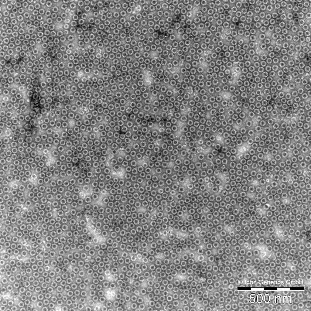
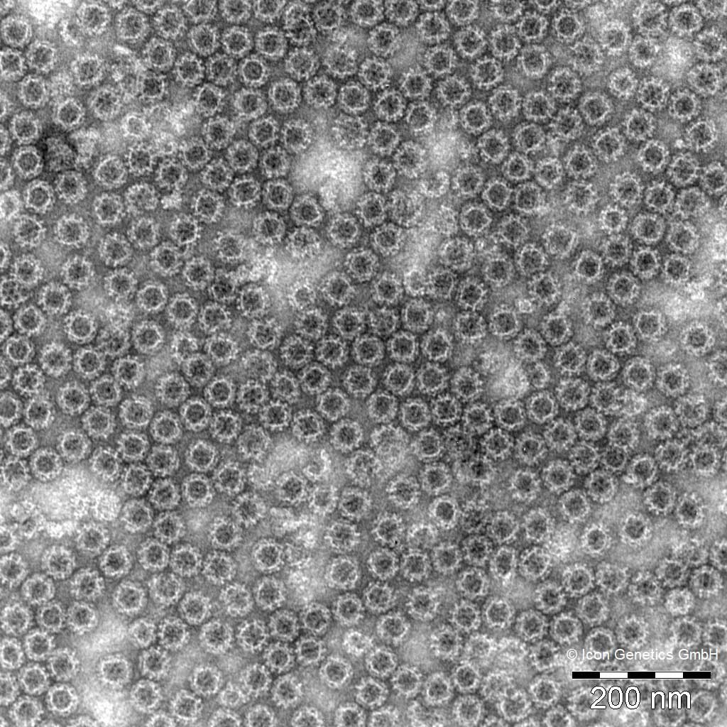
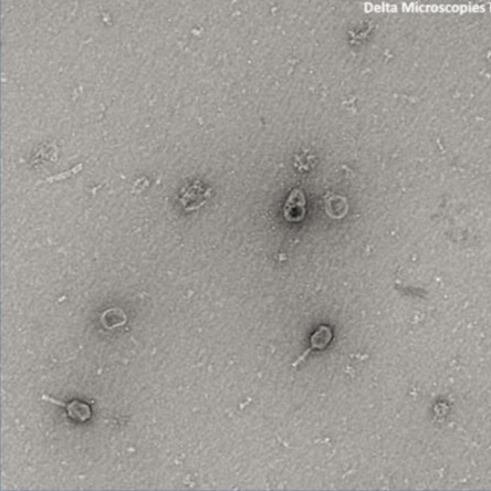
Negative staining with Uranyless examined by TEM

norovirus virus-like particles (VLPs)

UranyLess -Negative Staining UranyLess -Negative Staining

Electron micrographs of highly purified plant-produced
norovirus virus-like particles (VLPs) examined by
transmission electron microscopy (Zeiss EM900,
Carl Zeiss Microscopy, Jena, Germany). Samples
were collected on formvar coated cooper grids and
contrasted with UranyLess. Micrographs were taken
with a SM-1k-120 Variospeed slow scan camera
(TRS, Moorenweis, Germany) using the iTEM software
from Olympus SIS (Münster, Germany).

Thanks to Dr André DIESSNER)
Icon Genetics GmbH  – D-06120 Halle/Saale Germany