 |
 |
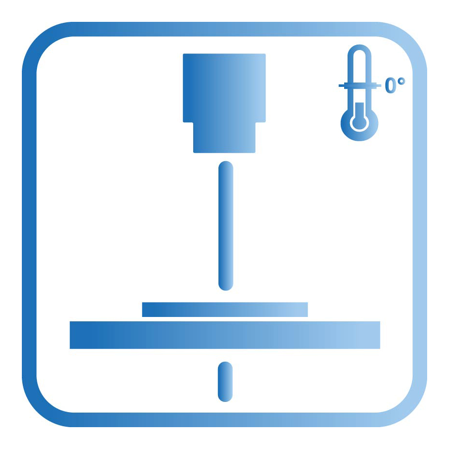 |
Processing at Cryo- or Room-Temperature
For Cryo-EM, vitrified samples can be prepared in different ways. In cryo-substitution, the samples are fixed at -90°C, then dehydrated and finally contrasted. After this freezing substitution, the sample is embedded in UV-polymerising plastic and can subjected to various processes, such as freeze fracture, freeze etching and heavy metal vapour deposition. To obtain a better ultrastructure of the sample, chemical treatment and embedding should be avoided. A pure vitrified cell or tissue sample can be cut in a cryo-ultramicrotome or milled by cryo-focused ion beam (FIB). This extremely fine milling procedure is done in a scanning electron microscope equipped with an ion source (gallium). The focused ion beam ablates the sample (which should not be thicker than 5-10µm) in a defined range down to 300-500nm. The FIB lamella created this way is largely artefact-free and can subsequently be visualised in cryo-TEM. Single particles such as viruses and molecular complexes can be directly imaged after vitrification.