NanoSuit Solution for SEM, aqueous Solution, 5ml
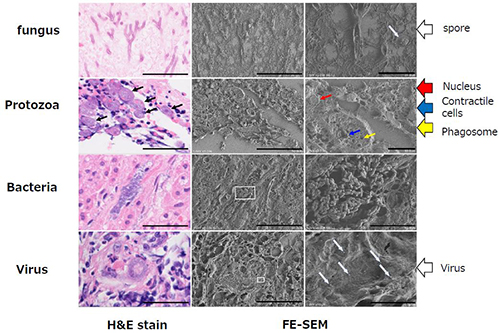
NanoSuit® is a novel technology to observe biological organisms, cells, bacteria, viruses or cellular compartments in a natural, living state using scanning electron microscopy (SEM). It forms a thin, conductive, moisture barrier layer between specimen and vacuum environment to hold moisture in the specimen. Simply drop the solution onto the object. No fixation or dehydration procedures needed.
Product Details
Description
Taking electron microscope images of biological objects in their natural state
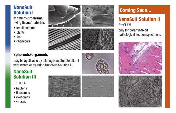
NanoSuit Selection Guide
NanoSuit Solution is available in two (soon three) types. Refer to the chart below to select the formulation which suits your application.
History
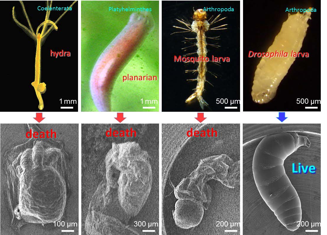
The aim of Professor Takahiko Hariyama was to investigate microorganisms using SEM, but due to loss of moisture under the vacuum condition, almost all organisms died in the electron microscope.
However, he discovered one kind of fly larva was able to survive under vacuum conditions.
He observed a very thin barrier layer was formed on the surface of the larva by electron beam irradiation. The barrier layer held moisture and kept the larva alive.
By mimicing this effect artificially, Hariyama and his colleagues invented the 'NanoSuit' technology.
Science
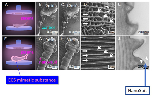
The 'NanoSuit'
- Aqueous solution of the biocompatible polymer. (biologically safe material)
- Just drop onto the objects.
- Polymerize by electron beam or plasma irradiation, then a thin barrier layer is formed on the surface of the objects.
- The barrier layer holds moisture contained in the objects and its conductivity provides clear SEM images.
Example (flower petal)
| Control | Control | |
|
|
Under vacuum, the petals are dried and do not retain original shape. | |
| Cryo-SEM | NanoSuit | |
 |
||
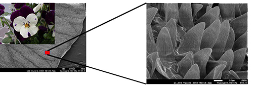
NanoSuit keeps the petal moist under vacuum and provides fine structure images of naturally shaped petal surface.
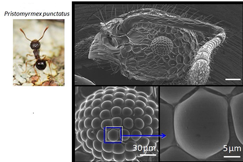
Example (insect)
Observation with NanoSuit of the minute structure of the insect's body surface with a simple operation.
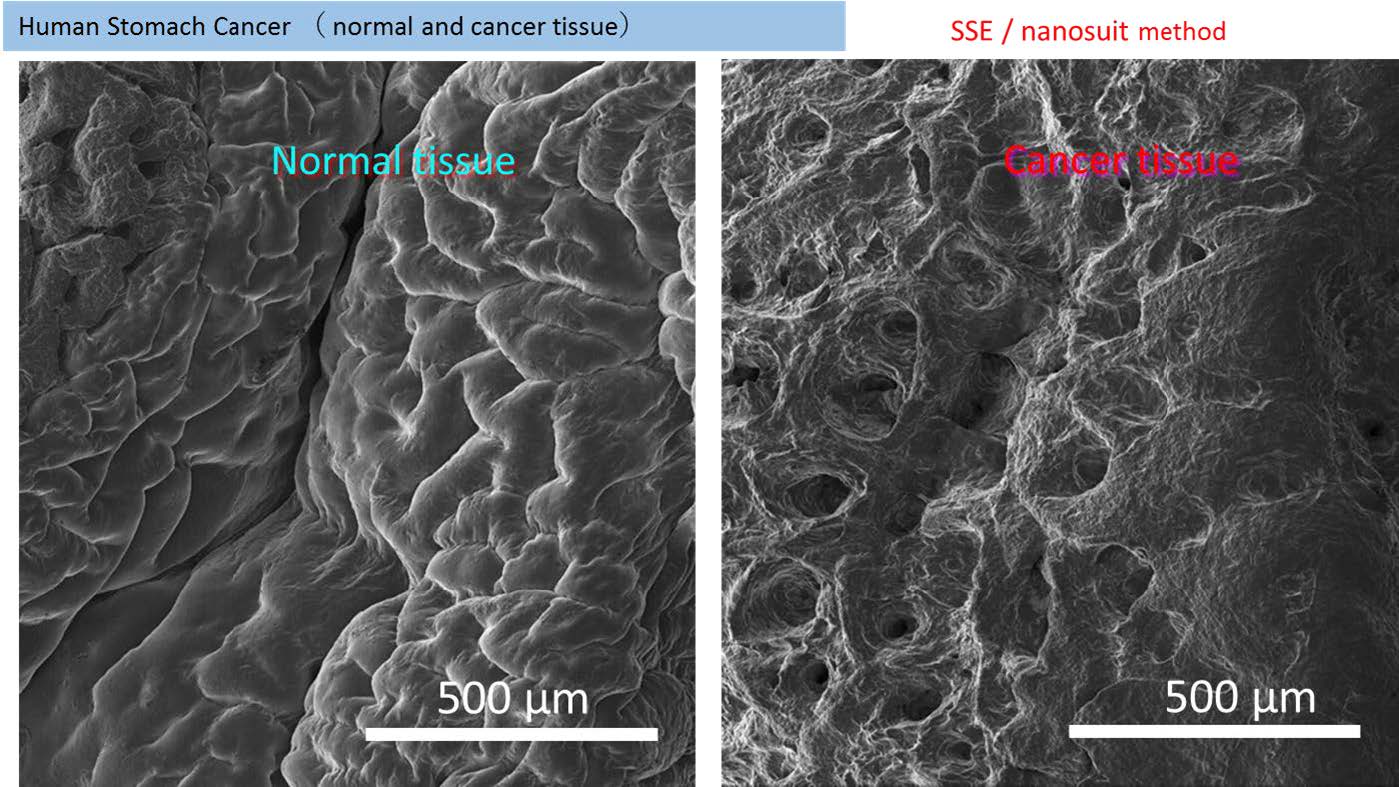
Example (living tissues)
NanoSuit keeps the texture of living cells and enables to distinguish between cancer tissue and normal tissue.
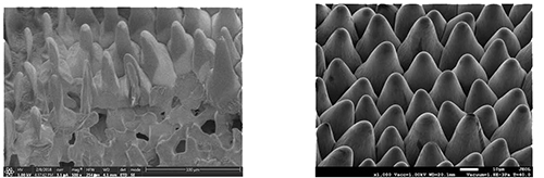
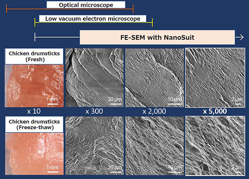
Example (chicken drumsticks)
Protection by NanoSuit solution of chicken drumsticks (fresh) surface. Observed by high resolution FE-SEM shows fibrous microstructures of uniform size.
On the other hand, chicken drumsticks (freeze-thaw) examined by the same method, show a broken fine structure of the surface and liquid substances seeping out to the surface.
Images suggest that such a difference in surface condition may be the cause for the dry feeling of the thawed food.
Example (CLEM; Correlative light electron microscopy)
By using the NanoSuit-CLEM method, observation of sample with a light and an electron microscope is possible. Since metal deposition is not required, samples can be stored on glass slides for optical microscopy. A lot of information can be obtained by combining the stained results from the light microscope with the 3-dimensional, high-magnification results from the electron microscope.
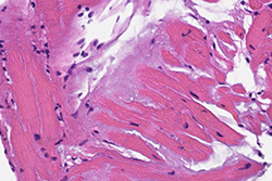
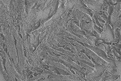
Example (pathological specimen)
Amyloid deposition of cardiac muscle tissue - NanoSuit-CLEM Method
By using the NanoSuit-CLEM method, it is possible to make a diagnosis by combining amyloid deposition in an image stained by light microscopy and changes in the surrounding tissue morphology observed using electron microscopy.
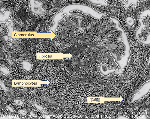
Example (pathological specimen - Nephritis)
The glomerulus is broken about half due to inflammation and lymphocytes are infiltrated.
References
Kawasaki, H., Itoh, T., Takaku, Y. et al. The NanoSuit method: a novel histological approach for examining paraffin sections in a nondestructive manner by correlative light and electron microscopy. Lab Invest 100, 161-173 (2020). https://doi.org/10.1038/s41374-019-0309-7
Shinmura K, Kawasaki H, Baba S, et al. Utility of Scanning Electron Microscopy Elemental Analysis Using the 'NanoSuit' Correlative Light and Electron Microscopy Method in the Diagnosis of Lanthanum Phosphate Deposition in the Esophagogastroduodenal Mucosa. Diagnostics (Basel). 2019;10(1):1. Published 2019 Dec 18. doi:10.3390/diagnostics10010001
Takaku Yasuharu, Suzuki Hiroshi, et al. 'NanoSuit®' preserves wet samples in high vacuum: direct observations on cells and tissues in field-emission scanning electron microscopy. R. Soc. open sci.4160887. 2017A modified.
Storage Conditions
| Short Term: | Refrigerator, 4°C |
| Long Term: | Frozen, -10°C |
NanoSuit Solution II for CLEM - Coming Soon!
More Information
| Application |
EM
SEM
|
|---|---|
| Shipping Class |
Reefer Cargo
|
| Working Field |
Scanning Electron Microscopy
|
Downloads

Who Viewed This Also Viewed