A little Story of Cell Counting
Cell counting is a standard procedure routinely used in (cell culture) laboratories, e.g. to maintain healthy cell cultures or before freezing cells. For many experiments the exact number of cells has to be known in order to e.g. adjust the amount of chemicals required, but also for the consistency and reproducibility of experiments. Furthermore, it is important to know the accurate cell count for protocols like transfection, cell proliferation studies or qPCR (quantitative PCR). Simultaneously, viability can be determined.
Originally developed for the determination of the blood count, glass counting chambers (hemocytometers) are used to identify the number of blood cells in a sample. Meanwhile many other counting chambers are available to count bacteria, viruses or other pathogens (in the blood).
Ways to count cells
The cell count can be obtained either by counting the cells manually in a counting chamber such as the traditional hemocytometer or by using automated methods like flow cytometers, spectrophotometers, coulter counters or cell counters that use an image analysis software.
A. Manual cell counting
B. Automated cell counting
A. Manual cell counting
 1) Counting chamber: For manual counting, a cell suspension is transferred into a special counting chamber (e.g. hemocytometer). A counting chamber is a glass/plastic slide into which a grid of a defined size is etched. The cells in several areas of the grid are then counted under the light microscope and the number of cells is determined by extrapolation to the total volume. If special dyes (e.g. trypan blue, which only stains dead cells blue) are added, the viability of the cells (i.e. the ratio of living cells to the total number of cells) can also be determined. Glass chambers can be reused indefinitely but only one or two samples can be counted at the same time. Plastic counting chambers are disposable (interesting e.g. for infectious material) and have two or 4 sample counting areas in one slide.
Application: routine cell counting for maintenance.
2) Plaque assay/CFU (colony forming unit) count on nutrient medium: If youwant to determine which subgroups of cells should survive a certain treatment (e.g. bacteria resistant to a given antibiotic) by choosing the growth conditions (media composition, oxygen content, etc...), the plaque assay is suitable. For this purpose, a highly diluted cell suspension is spread evenly on solid growth medium in a Petri dish. The cell colonies that emerge after about 12-14 hours are then counted manually. Since only the surviving cells form a colony this test doesn´t give any indication about viability in the starter cells.
Application: drug screening.
| Advantages of manual cell counting | Disadvantages of manual cell counting |
| Relatively fast | Cells may need to be diluted (less accurate) |
| Inexpensive, sustainable (when glass chamber is being used) | Adherent cells must be detached and well suspended (possibly disrupting the experimental conditions, affecting viability) |
| Cell number and viability at the same time | 1-4 samples only per chamber |
| Cells can be seen (visual inspection) | Subjective errors possible |
interesting products
B. Automated cell counting
1) Coulter counter (volume measurement of cells): A Coulter counter is measuring the change in electrical resistance in a solution through the cells it contains. Apart from counting particles (not just cells), a Coulter counter can measure their size, as well. Additionally, this method is not as expensive as flow cytometers making it all in all the method of choice for cell cycle experiments.
Application: method of choice for cell cycle experiments.
2) Spectrophotometry: Measures the absorbance of light/optical density (OD) in a cell suspension. The higher the cell concentration, the cloudier the cell suspension in which the cells grow. This method is used e.g. to check bacterial cultures for their growth.
Application: Growth monitoring in bacterial cultures.
3) Flow cytometry: In a flow cytometer cells in a solution flow individually through a laser beam causing a light scatter (or in case fluorescence labelled cells are used a fluorescence signal). A light detector collects and analyses the backscattered light/fluorescence thereby picking up not only the cell count but also differences in form, structure and color of the cells.
Application: sophisticated method for cell analyses.
 4) Image Analysis: Nowadays an increasingly large number of devices are available that count cells via an image analysis software. Many also have additional features that allow researchers to characterize cells further via the analysis of viability, cell size, cell cycle, fluorescence expression and apoptosis.
Many microscopes now are equipped with an AI-controlled imaging software that makes cell counting intuitive, fast and effective for the user (e.g. the AI cell counting module of the Labscope software for the Zeiss Axiovert, Primovert and Primo Star microscopes).
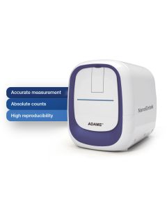
ADAMTM-MC2 is a fast and accurate automated fluorescent cell counter
interesting products
-
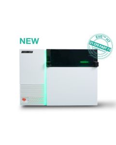
 Art.Nr: NE-EVE-HT-FL
The EVE™ HT FL is a high-throughput automated fluorescence cell counter equipped with bright field and two fluorescence channels, UV for DAPI and blue for Acridin Orange (AO) detection. In only 3 minutes, up to 48 samples can be counted and analyzed. EVE™ HT FL delivers highest precision and accuracy for both cell lines and primary cell analysis in a variety of applications.
-
 Art.Nr: NE-ADAM-MC-Plus
ADAM stands for Advanced Detection and Accurate Measurement. The ADAM™ MC Plus is the new standard of highly accurate automated fluorescence cell counting of primary mammalian cells and cell lines. It is equipped with bright field and two fluorescence channels (AO/DAPI) for sensitive fluorescence dye detection. LED optics and CMOS detection technologies make analysis more accurate and reliable. It measures the number of total cells, viable and non-viable cells and calculates the cell viability. In addition, it analyzes the cell size and aggregation ratio.
-
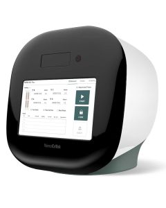
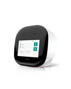
 Art.Nr: NE-ADAMII-LSADAMII™ LS is an accurate and easy-to-use bench-top system for fluorescent cell analysis. It enables performing of various cell assays with up to 4 channels (bright-field, DAPI, GFP, RFP), including cell counting, viability, fluorescent expression (transfection efficiency), apoptosis and cell cycle histograms.
 -
 Art.Nr: NE-ADAM-MC2The ADAM MC2 is an automated, easy-to-use and cost-efficient fluorescence cell counter. It operates with disposable microfluidic chips and analyses PI- or AO/PI-stained cell nuclei. LED illumination and CMOS detector technology enables precision analysis with reliable results in your lab.
 -
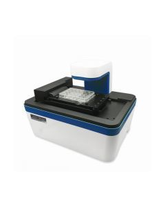 Art.Nr: NE-JS1000S
- Multi-channel fluorescence imaging
- Compact and compatible with standard CO2 incubators
- Fully automated X-Y-Z stage
- Easy and powerful software
- Capture and analyze images in real time
-
 Art.Nr: NE-EVE-MC
The EVE™ automatic cell counter uses state-of-the-art optics and image analysis to automate cell counting. EVE™ is a bench-top counter, designed to measure cell count and viability (live, dead and total cells) accurately and precisely, using the standard trypan blue technique.
EVE™ is the succeeding model of the established Countess® automated Cell Counter.
-
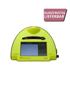
 Art.Nr: NE-EVE-HT
The EVE-HT is a high-throughput automated cell counter ideal for large scale cell production or development. High efficiency, low sample volume and a speed of 48 samples in only 3 minutes enable time saving reliable workflows. Cell declustering, counting and viability measurement with up to 48 channels simultaneously enable large scale projects with high precision.
-
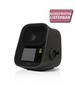 €3,900.00 €4,641.00Art.Nr: NE-EVE-MC2
EVE™ PLUS is the 2nd generation of EVE™, an image based automated cell counter. The EVE™ PLUS is a benchtop automated cell counter that performs cell count and viability measurements using trypan blue//erythrosin B solution. It is comparable to the nucleus counting method with PI (Propidium Iodide) for all cell lines with accuracy and precision. EVE™ Plus also identifies and counts cell agglomertes as several individual cells for accurate analysis.
- Less than 1 s for cell counting (world´s fastest automated cell counter)
- NEW: Autofocus (< 10 s for cell counting)
- Easy to use
- More accurate by improved hardware and counting algorithm
€3,900.00 €4,641.00 -
Starting at €78.00 €92.82
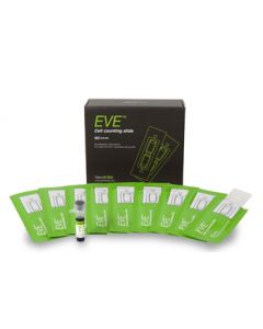
The EVE™ cell counting slides contain two chambers. This allows measurement of two different samples or to perform a reference test of the same sample. Each kit includes 1,5ml Trypan Blue Solution (0,4%) per 50 slides. The kit is fully compatible with Countess® automated cell counters.
Starting at €78.00 €92.82