Array Tomography
(4)Array Tomography is one way to acquire 3D data series of ultra or semi-thin sections of a tissue sample. Thereby, the sample is cut with an ultramicrotome and the sections are collected on a substrate for subsequent imaging with a SEM. Depending on the research project more or less sections are required to completely reconstruct a region of interest.
In comparison to Serial Block-Face Scanning EM (SBFSEM) and Focused Ion Beam (FIB), Array Tomography is a non-destructive technique. The collected sections can be stored, re-imaged at difference regions and resolutions, re-stained with heavy metals to enhance contrast or with fluorescent markers for correlative light and electron microscopy (CLEM). By now, Array Tomography is not only used for Connectomics but also in other fields of research, e.g. single cell reconstructions or even to gain 3D data of complex materials.
We offer different automated and manually controlled tools to collect serial sections depending on their amount, size and thickness:
-
 Art.Nr: R75850
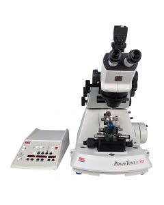
Computer-controlled PT3D with video display from the RMC PowerTome series. The PT3D features an unique advance of 1000µm for uninterrupted sectioning of ultra-thin sectons. It has been developed for 3D reconstruction work, array tomography or applications where high amounts of trimming are necessary such as cryo-sectioning. It is also equipped with the patented PowerDrive® technology for the preparation of ultra- and semi-thin sections for electron microscopy. The mechanical stroke force is particularly advantageous for cutting hard and large specimen.
-
 Art.Nr: R-ATUMtome
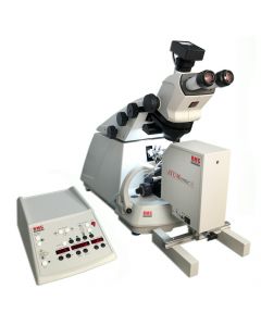
The ATUMtome by RMC, a tape collecting ultramicrotome with automated section collector. Initially developed at Harvard University in the lab of Jeff Lichtman. The ATUMtome is a unique advanced system for ultrathin sectioning of resin-embedded specimens and collecting sections on tape for SEM imaging and subsequent 3-D reconstruction (Array Tomography).
-
Starting at
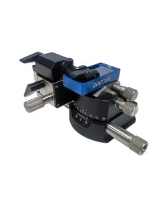
The ASH2 is used to efficiently collect and align hundreds of serial sections onto a variety of substrates for subsequent imaging in LM and SEM. The advanced substrate holder ASH2 quickly mounts onto the knife stage of RMC PowerTome ultramicrotomes.
Starting at
-
Starting at
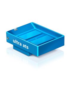
The DiATOME ultra ats was developed for mounting series of ultra-thin sections directly on (conductive) substrates such as silicon wafers or ITO slides. Using Array Tomography 3D data sets of the section series can be recorded by subsequent SEM imaging. The large knife boat is equipped with a water outlet at the bottom.Starting at


Array Tomography
Connectomics – the map of neuronal pathways and connections of an organism’s nervous system – has been the driving force for the development of 3-dimensional (3D)-imaging techniques in light and electron microscopy. Even the latest fluorescence imaging approaches, such as super resolution microscopy (STED, PALM, STORM) lack the resolution necessary for a detailed reconstruction of neuronal systems including synaptic connections.
Due to resolution limits of light microscopy researchers started to investigate in techniques capable of reconstructing neuronal system at sufficient resolution to display neuronal connectivity. Within the last decade three powerful techniques based on scanning electron microscopy (SEM) have been developed and tailored for volume imaging at nanometer resolution: Serial Block-Face Scanning EM (SBFSEM), Array Tomography (AT) and Focused Ion Beam (FIB).
Array Tomography
Array Tomography is one way to acquire 3D data series of ultra or semi-thin sections of a tissue sample. Thereby, the sample is cut with an ultramicrotome and the sections are collected on a substrate for subsequent imaging with a SEM. Depending on the research project more or less sections are required to completely reconstruct a region of interest.
In comparison to Serial Block-Face Scanning EM (SBFSEM) and Focused Ion Beam (FIB), Array Tomography is a non-destructive technique. The collected sections can be stored, re-imaged at difference regions and resolutions, re-stained with heavy metals to enhance contrast or with fluorescent markers for correlative light and electron microscopy (CLEM). By now, Array Tomography is not only used for Connectomics but also in other fields of research, e.g. single cell reconstructions or even to gain 3D data of complex materials.
References
Q&A: Array tomography. Smith SJ; BMC Biol. 2018 Sep 6;16(1):98.
A targeted 3D EM and correlative microscopy method using SEM array tomography. Burel et al; Development. 2018 Jun 21;145(12).
Multimodal Hierarchical Imaging of Serial Sections for Finding Specific Cellular Targets within Large Volumes. Wacker et al; J Vis Exp. 2018 Mar 20; (133). Video-Link.
A carbon nanotube tape for serial-section electron microscopy of brain ultrastructure. Kubota et al; Nat Commun. 2018 Jan 30;9(1):437.
Hierarchical imaging: a new concept for targeted imaging of large volumes from cells to tissues. Wacker et al. BMC Cell Biol. 2016; 17: 38.
High-resolution whole-brain staining for electron microscopic circuit reconstruction. Mikula and Denk; Nat Methods. 2015 Jun;12(6):541-6.
New developments in electron microscopy for serial image acquisition of neuronal profiles. Kubota Y; Microscopy (Oxf). 2015 Feb;64(1):27-36.
Array tomography. Wacker I and Schroeder RR; J Microsc. 2013 Nov;252(2):93-9.