GraFIX, a method for single particle cryo-electron microscopy (cryo-EM)
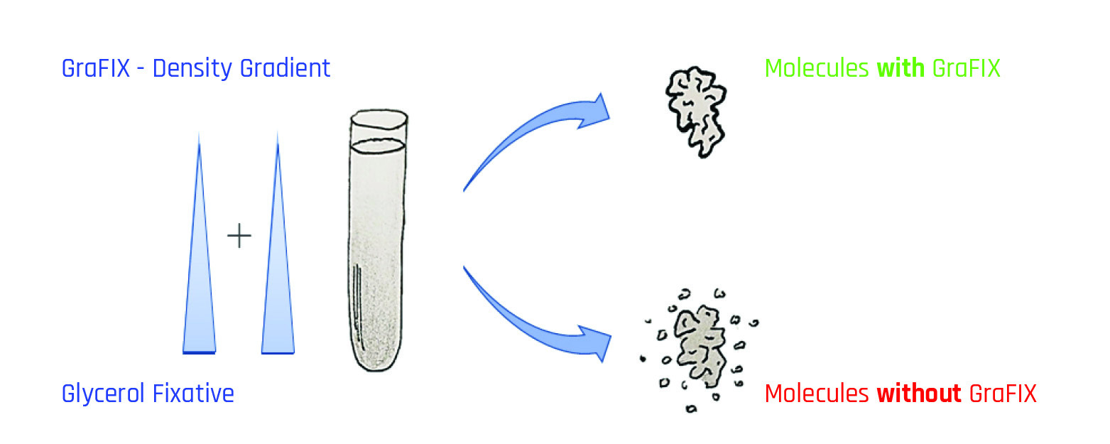
Fig. 1.: GraFIX method: The centrifugation tube contains a gradient of glycerol or sucrose and cross-linking reagent (e.g. glutaraldehyde or formaldehyde). Separation is based on sedimentation speed (rate zonal centrifugation). The GraFix treatment improves the properties and stability of the molecular complexes in contrast to the untreated sample, which shows degradation and destabilization artifacts.
For single particle structure determination in cryo-EM, purified proteins are frozen (vitrified) in their native state. However, the imaging contrast in cryo EM is very weak due to the low density of the elements of life. Negative staining can be used to increase contrast, but unstained native structural analysis is the method of choice. During macromolecule purification, sample heterogeneity also often occurs because of degradation processes and protein complexes are destabilized by in vitro buffer solutions. These and other complications can seriously compromise high-resolution 3D reconstruction.
The GraFix method (gradient fixation) can dramatically increase the quality and stability of proteins for single particle analysis (Kastner et al., 2008; Stark et al. 2010). Usually, macromolecules are purified and concentrated by density gradients. This is exactly where the GraFix method steps in. During rate-zonal density gradient ultracentrifugation, the macromolecules are exposed to a low-dose chemical crosslinking agent (fixative). This is added directly during the preparation of the gradient (Fig. 1).
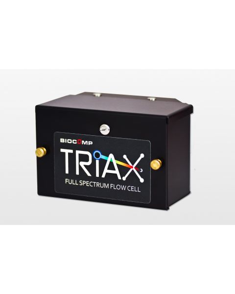
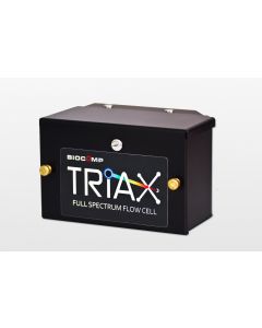
Density gradients can be prepared in a few minutes using the tilted-rotation method with a Gradient Master or a Gradient Station from BioComp Instruments, Canada. Linear gradients with increasing concentration of e.g. glycerol or sucrose and an increasing content of fixative are produced. The molecules pass through this density gradient during ultracentrifugation, where they are separated based on their sedimentation speed. At the end of ultracentrifugation, bands of purified molecules that have undergone gentle fixation are formed. The concentrated sample can then be removed from the tubes, e.g. manually with a cannula. For higher purity and reproducibility, the sample can be collected with a BioComp Fractionator or a Gradient Station. The sample can be analyzed directly during fractionation by UV absorption or fluorescence emission with a TRIAX flow cell.
After fractionation, the individual particles can be additionally contrasted by negative staining or analyzed directly in cryo-EM after a short buffer exchange without staining.
Several studies have shown that the GraFix method significantly improves the imaging quality of single particle properties as well as molecular dispersion in cryo-EM (Stark et al. 2010; Wang et al. 2022). GraFix is highly reproducible and suitable for routine use.
Kastner B. et al. 2008, Nat Methods “GraFix: sample preparation for single-particle electron cryomicroscopy.”
Stark H. et al. 2010, Methods Enzymol “GraFix: stabilization of fragile macromolecular complexes for single particle cryo-EM.”
Wang L. et al. 2022, Nat Commun “Structure of nucleosome-bound human PBAF complex”
PRODUCTS
-
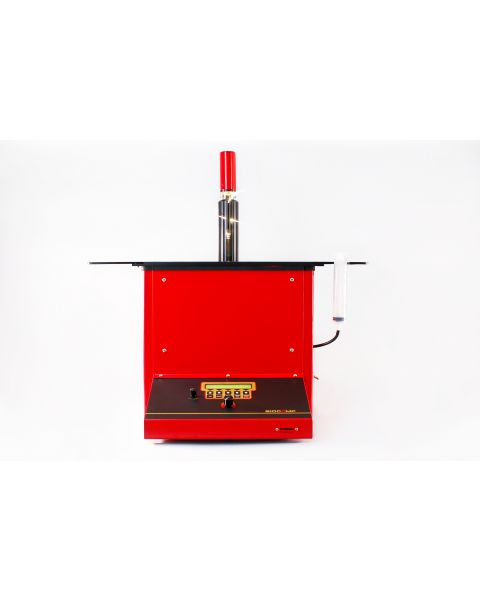
 Art.Nr: B153-002Base unit for generating linear density gradients and fractionation of centrifuged samples from the ultracentrifuge tube. The tube remains undamaged. The Gradient Station™ combines the Piston Gradient Fractionator™ and the Gradient Master™ in one device. All technical data, parts and accessories are identical to those of each instrument. Tube holders are required for each tube size. These must be purchased in addition to the basic device. Get in touch with us and we'll find the best configuration for your application.
 -
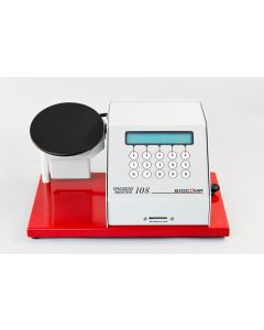 Art.Nr: B108-2Gradient Master™ Base Unit for making 6 identical linear gradients within minutes. MagnaBase™ Tube Holders are not included.
 -
 Art.Nr: B152-002Base unit for the fractionation of centrifuged samples in linear density gradients from ultracentrifuge tubes. The process is very reproducible and highest sample purity can be achieved. The tube remains undamaged. Separate Tube holders are required for each tube size. These must be purchased in addition to the basic device. Get in touch with us and we'll find the best configuration for your application.
 -
 Art.Nr: BFC-1
C-1 is the single wavelength version. The flow cell can be configured to record UV or fluorescence, allowing overlay of separate UV and VIS scans. Wavelength and fluorophore changes are made by changing the LEDs, photodiodes and filters.
-
 Art.Nr: BFC-2
FC-2 allows two input wavelengths so the UV and VIS scans are obtained in the same gradient. Another use is the dual UV scan at 260 and 280 nm, providing a 260/280 ratio that reflects the change in nucleotide/protein composition of a particle.
-
 Art.Nr: BFC-3
FC-3 is used for three wavelength scans, one or two for UV scans and the other one or two for the VIS scans. Up to three wavelengths can be recorded simultaneously in the same gradient. All three LEDs are driven and tuned simultaneously by using the Triax™ software.